
Berperan sebagai salah satu Presenter di Seminar Internasional 2nd Borobudur International Symposium 18 November 2020 di Universitas Muhammadiyah Magelang, Indonesia.
Membahas menenai penelitian terbaru yang sedang ramai didiskusikan di antara para ilmuan
Berperan sebagai salah satu Presenter di Seminar Internasional 2nd Borobudur International Symposium 18 November 2020 di Universitas Muhammadiyah Magelang, Indonesia.


Andi Hasad dkk Mendapatkan HAKI (Hak Kekayaan Intelektual) Dari Hasil Penelitian Strategi Nasional Institusi (PSNI), Biaya Dari Ristek Dikti Tahun 2018
Alhamdulillah, Penelitian Strategis Nasional Institusi (PSNI) tahap lanjutan / tahun ke-2, lolos pendanaan KemenRistek Dikti Direktorat Jenderal Penguatan Riset Dan Pengembangan. Semoga pelaksanaannya dimudahkan dan dilancarkan, hasilnya bermanfaat bagi peneliti/instansi, masyarakat, bangsa dan negara.
Perguruan Tinggi : Universitas Islam 45 Bekasi (Unisma Bekasi)
Skema : Penelitian Strategis Nasional Institusi (PSNI)
Nama Ketua Peneliti : Andi Hasad
Status Usulan : Lanjutan
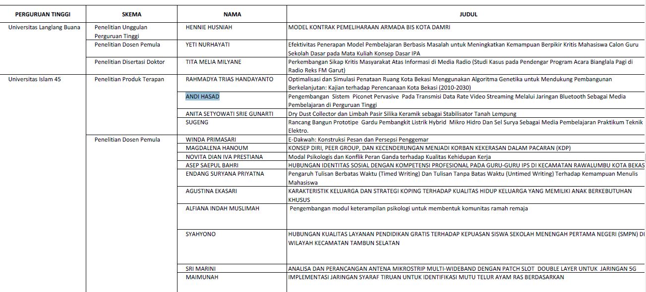
Judul : Pengembangan Sistem Piconet Pervasive Pada Transmisi Data Rate Video Streaming Melalui Jaringan Bluetooth Sebagai Media Pembelajaran di Perguruan Tinggi
Dalam pendanaan tahun 2018, tercatat 4 dosen Peneliti (semua dari Fakultas Teknik Unisma Bekasi) yang lolos pendanaan skema Penelitian Strategis Nasional Institusi (PSNI) untuk tahap lanjutan / tahun ke-2, sedangkan dalam skema Penelitian lainnya untuk tahap baru terdiri dari 19 Peneliti untuk skema Dosen Pemula (PDP), 3 Peneliti skema Penelitian Disertasi Doktor (PDD) dan 1 Peneliti untuk skema PKPT , dari berbagai Fakultas di Universitas Islam 45 Bekasi.

Selengkapnya surat dan daftar Penerima Pendanaan Penelitian dan Pengabdian Masyarakat di Perguruan Tinggi Tahun 2018, dapat dilihat pada lampiran file berikut :
Referensi:


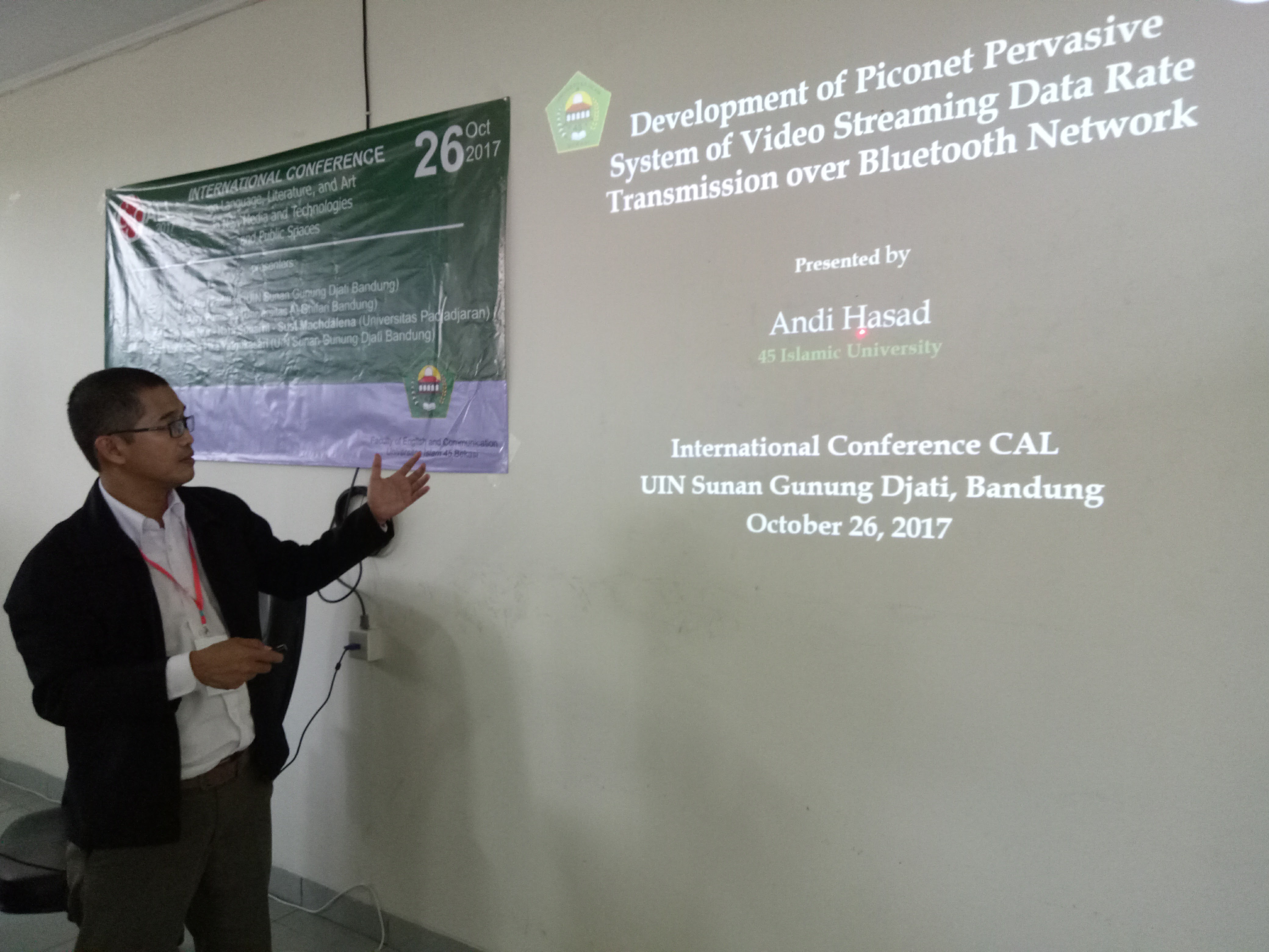
Presentasi di International Conference CAL (IC-CAL) 26 Oktober 2017 di UIN Sunan Gunung Djati, Bandung
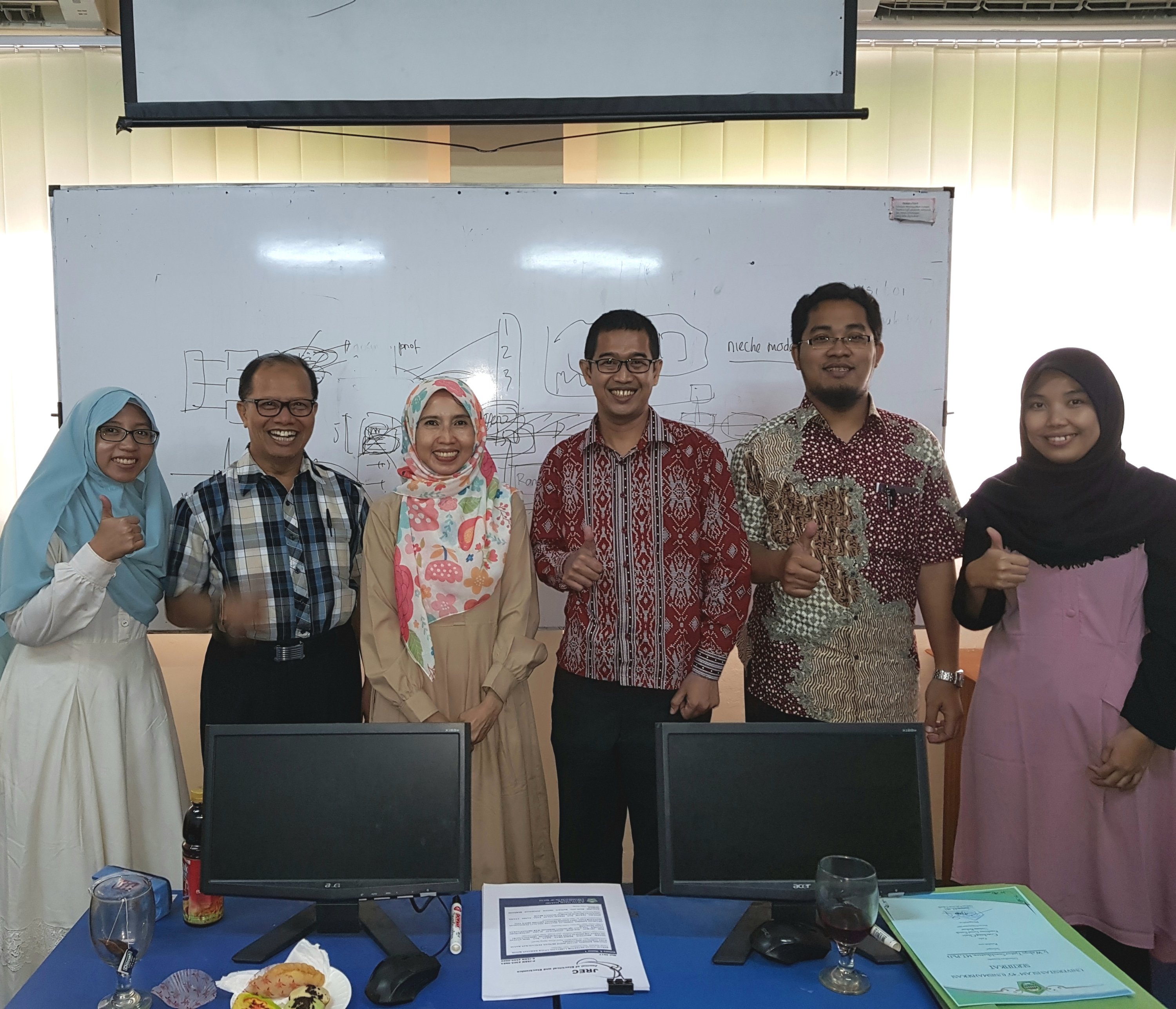


Coaching clinic pengelolaan jurnal program studi Teknik Elektro dan Teknik Elektronika di Sainstech Unisma Bekasi.
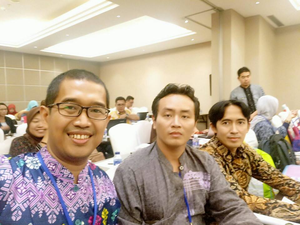

Seminar Hasil Penelitian DRPM Kemenristek Dikti Pendanaan 2016
Alhamdulillah, Penelitian Produk Terapan (PPT) lolos pendanaan KemenRistek Dikti Direktorat Jenderal Penguatan Riset Dan Pengembangan. Semoga pelaksanaannya dimudahkan dan dilancarkan, hasilnya bermanfaat bagi peneliti/instansi dan masyarakat.
Perguruan Tinggi : Universitas Islam 45 Bekasi (Unisma Bekasi)
Skema : Penelitian Produk Terapan
Nama : Andi Hasad
Judul : Pengembangan Sistem Piconet Pervasive Pada Transmisi Data Rate Video Streaming Melalui Jaringan Bluetooth Sebagai Media Pembelajaran di Perguruan Tinggi
Dalam pendaanaan tahun 2017, tercatat 4 dosen (semua dari Fakultas Teknik Unisma Bekasi) yang lolos pendanaan skema Penelitian Produk Terapan (PPT), sedangkan dalam skema Penelitian Dosen Pemula (PDP), terdapat 10 dosen dari berbagai Fakultas yang dinyatakan lolos pendanaan. Selengkapnya sebagai berikut:

Penerima Pendanaan Penelitian Dan Pengabdian Masyarakat Di Perguruan Tinggi Tahun 2017. Kementerian Riset, Teknologi, Dan Pendidikan Tinggi Direktorat Jenderal Penguatan Riset Dan Pengembangan.
Download surat resmi Sumber Simlitabmas Ristek Dikti

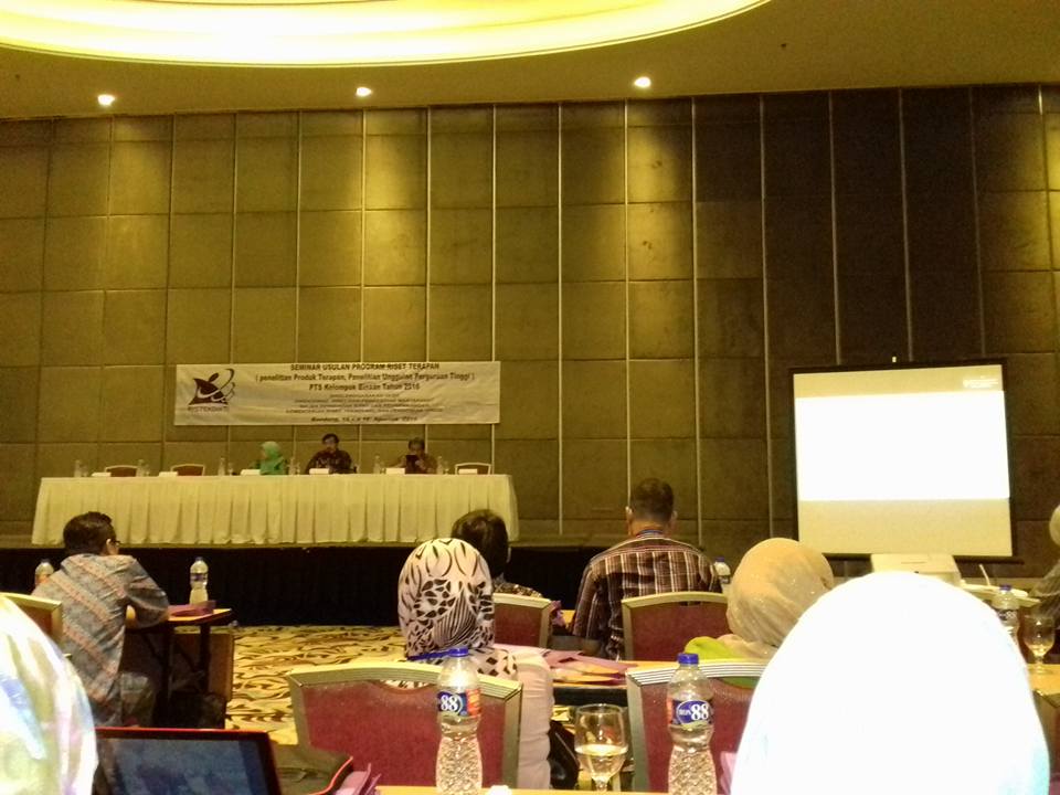

Kegiatan Seminar Usulan Program Riset Terapan ( Penelitian Produk Terapan, Penelitian Unggulan Perguruan Tinggi) Tahun 2016 ini bertujuan antara lain:
Dari Universitas Islam 45 Bekasi, ada 4 proposal yang dinyatakan lulus Desk Evaluation, dan masuk ke tahap berikutnya presentasi seminar usul penelitian, yaitu:

Salah satu reviewer Dikti pada kegiatan ini adalah Prof. Dr. Kuncoro, S.T., M.T., Guru Besar Universitas Sebelas Maret, Solo. Kegiatan ini berjalan lancar dan sukses , 15-16 Agustus 2016, berlokasi di Hotel Harris, Jl. Peta Bandung.
Hasil dari kegiatan ini merupakan pertimbangan bagi Direktorat Riset dan Pengabdian Masyarakat, Direktorat Jenderal Penguatan Riset dan Pengembangan, dalam menentukan usulan baru penelitian yang akan didanai pada tahun anggaran 2017.
Lampiran:
Undangan Peserta Seminar Usulan Penelitian Riset Terapan Tahun 2016 di Bandung
Lampiran Undangan Seminar Usul Riset Terapan
Sumber:
Panduan Kegiatan Seminar Susulan Program Riset Terapan ( Penelitian Produk Terapan, Penelitian Unggulan Perguruan Tinggi) PTS Kelompok Binaan. DRPM, Ditjen Penguatan Riset dan Pengembangan. Ristek DIKTI, 2016.
Mesothelioma: Do asbestos and carbon nanotubes pose the same health risk?. Carbon nanotubes (CNTs), the product of new technology, may be used in a wide range of applications. Because they present similarities to asbestos fibres in terms of their shape and size, it is legitimate to raise the question of their safety for human health. Recent animal and cellular studies suggest that CNTs elicit tissue and cell responses similar to those observed with asbestos fibres, which increases concern about the adverse biological effects of CNTs. While asbestos fibres’ mechanisms of action are not fully understood, sufficient results are available to develop hypotheses about the significant factors underlying their damaging effects. This review will summarize the current state of knowledge about the biological effects of CNTs and will discuss to what extent they present similarities to those of asbestos fibres. Finally, the characteristics of asbestos known to be associated with toxicity will be analyzed to address the possible impact of CNTs.
Introduction
Carbon nanotubes (CNTs) have unique chemical and physical characteristics as a result of their nanostructure. CNTs may be used in a wide range of applications, in fields as diverse as electronics and medicine. Due to their widespread use, it is important to determine the safety of CNTs for the protection of ecological systems and human health. Research to investigate the biological effects of CNTs is advancing today in order to foresee and prevent their potentially harmful effects. CNTs have fibrelike characteristics in terms of their elongated shape, dimensions and aspect ratio. As particles with at least one dimension of less than 100 nm, they correspond to High Aspect Ratio Nanoparticles (HARN) . In light of the health impact of mineral fibres, especially the fibrogenic and carcinogenic potency of asbestos fibres, and the health and socio-economical tragedies caused by unregulated asbestos utilization, the increasing development and uses of CNTs have triggered concern about their potential toxicity. In recent years, several publications have reported the effects of CNTs. Most studies have concerned animal and cell responses, focusing primarily on respiratory diseases, especially the inflammatory effects in the lung. However, while inhalation is one important probable route of contamination, it must be kept in mind that there are other relevant routes of exposure. A severe primary cancer, malignant mesothelioma (MM), has been closely linked to asbestos exposure . Epidemiological and animal studies have shown that asbestos fibres are not the only fibres to be associated with a risk of MM development. Epidemiological studies have demonstrated a higher incidence of MM in populations exposed to asbestiform and non-asbestos fibres. Some manmade vitreous fibres have caused MM in animal experiments. The question of whether CNTs might potentially be linked to MM development justifies further research in this area. Moreover, on the basis of the literature, CNTs have already shown effects in animals and in cell systems that are similar to those observed with asbestos fibres. Two recent studies showed the occurrence of MM in genetically-modified cancer-sensitized mice and in conventional Fischer 344 rats exposed to CNTs by intraperitoneal or intrascrotal administration respectively. These initial results underline the urgent need for information to further our knowledge about CNTs’ potential to cause MM.
MM is a primary tumour of the serosas caused by the neoplastic transformation of mesothelial cells. In populations exposed to asbestos fibres, MM mainly occurs in the pleura, and to a lesser extent in the peritoneum and pericardium. MM is considered to be highly specific to asbestos exposure, and is found in from 60% to over 80% of cases. In France, the calculated risk of MM attributable to occupational asbestos exposure was estimated at 83.2% (95% CI 76.8 to 89.6) in men, and 38.4% (95% CI 26.8 to 50.0) in women. Many studies carried out to investigate pleural and mesothelial cell response to asbestos fibres have made it possible to reach sound hypotheses about the mechanism of action of asbestos fibres in neoplastic mesothelial cell transformation.
The aim of the present review is to explore whether our knowledge of the mechanism of action of asbestos fibres could offer a useful paradigm to provide a warning or predict the risk of CNTs, to interpret data on animal and cellular responses, and to evaluate their potential health effects. For the purposes of our discussion, we consider three points: (i) the fate of asbestos fibres following exposure; (ii) their effects on mesothelial cells and the biological mechanism associated with the cell response; (iii) the nature of the fibre parameters involved in the harmful effects, and their similarities with CNT characteristics. We begin with a summary of current knowledge on the toxicology of CNTs, then look at asbestos fibres’ mechanisms of action, focusing on carcinogenic effects at the pleural level. Finally, we address the similarities between asbestos and CNTs.
Toxicology of CNTs
Context of toxicological studies on CNT
Various kinds of CNTS have been the focus of toxicological studies. CNTs are heterogeneous in terms of their structure, impurities and physico-chemical properties. Both single-walled (SWCNTs) and multi-walled (MWC- NTs) CNTs have been examined in toxicological studies, including commercial and laboratory-made CNTs, whether purified or used as produced. The effects of CNTs have been investigated following in vivo exposure of rodents, and on several types of cells in culture. Most studies concerned pulmonary toxicity . Animal experiments mainly focused on inflammatory responses after exposure by intratracheal instillation or aspiration, or intraperitoneal injection. In vitro cell systems with several types of mammalian cells have been used to study inflammatory responses and genotoxicity. A few in vivo and in vitro studies were related to dermal toxicity, and some in vitro studies focused on neurons. Toxicity test systems on procaryotes were also used to assess genotoxicity. Here our focus will be on respiratory effects.

Biological effects of CNTs
Translocation
Biodistribution of CNTs after deposition in the lung or via other routes has been poorly investigated. A translocation of SWCNTs in various organs has been reported by several authors. In a recent study, MWCNTs deposited by intratracheal instillation in rats revealed clearance due to macrophage uptake and the lymphatic system without evidence of crossing the pulmonary barrier, six months after instillation. It can be noted that macrophage and lymphatic clearance was also demonstrated following administration or exposure to asbestos fibres. Erdely et al. suggest that the release of soluble inflammatory factors could circulate to the vascular blood compartment after lung deposition of CNTs. The release of circulating factors must be taken into consideration to account for fibre effects. While asbestos fibres have been detected in the pleura, soluble molecules could also account for the pleural response , and genotoxicity may be due to clastogenic factors. Additional studies are needed to determine the pharmacokinetics of CNTs. Regarding the numerous varieties of CNTs associated with a broad scale of physical and physico-chemical properties, fundamental studies will be necessary to establish the parameters leading the translocation process. Biological effects on mesothelial cells In vivo effects on mesothelial cells Six recently-published studies concerned CNTs’ effects on mesothelial cells. Three reported findings from animal experiments and three from cell system studies. One animal experiment concerned the mesothelial cell inflammatory response and pathological changes after intraperitoneal injection. The authors exposed C57Bl/6 mice to four samples of MWCNTs of different sizes and aggregation states. There was one sample of “short” MWC-NTs (from NanoLab, Inc; mean diameter: 14.8 ± 0.5 nm; mean length: 1–5 μm); two samples of “long” MWCNTs (Long1, from Mitsui & Co.; mean diameter: 84.9 ± 1.9 nm; mean length: 40–50 μm [24% > 15 μm of length]; Long2 from Univ. Manchester; mean diameter: 165 ± 4.7 nm; mean length: 20–100 μm [84% > 15 μm of length]); and one sample of more tangled MWCNTs (from NanoLab, Inc.; mean diameter: 10.4 ± 0.3 nm; mean length: 5–20 μm), as well as carbon black. At the same time, two samples of amosite fibres were tested; these were short fibres (4.5% > 15 μm of length) and long fibres (50.4% > 15 μm of length) known to be differently pathogenic in rodents. In prior experiments, inhalation and intraperitoneal exposure in rats to long amosite fibres revealed greater pathogenicity than short fibres in terms of fibrosis and cancer. In the study reported by Poland et al., inflammation was assessed after injection of 50 μg of MWCNTs/mouse, after 24 h and seven days. The end points were quantification of inflammation in peritoneal lavage and histology of diaphragm. Only long samples of MWCNTs and of amosite produced inflammation and granulomas. Histological analyses revealed the occurrence of “frustrated phagocytosis” by macrophages. These results thus demonstrated some similarities between the responses to the long forms of amosite and MWCNTs. Several of the effects of asbestos were also found with CNTs. There were higher inflammatory responses with samples of long fibres. Only the samples that contained long fibres caused granulomas and “frustrated phagocytosis”.
(Marie-Claude F Jaurand*1,2 , Annie Renier1,2 and Julien Daubriac1,2 Address: 1 INSERM, U674, Fondation Jean Dausset – CEPH, Paris, F-75010, France and 2 Université Paris 7, Paris, F-75013, France)
 Nanotechnology is developing rapidly throughout the world and the production of novel man-made nanoparticles is increasing, it is therefore of concern that nanomaterials have the potential to affect human health. The purpose of this study was to investigate the effects of maternal exposure to nano-sized anatase titanium dioxide (TiO2 ) on gene expression in the brain during the developmental period using cDNA microarray analysis combined with Gene Ontology (GO) and Medical Subject Headings (MeSH) terms information.
Results: Analysis of gene expression using GO terms indicated that expression levels of genes associated with apoptosis were altered in the brain of newborn pups, and those associated with brain development were altered in early age. The genes associated with response to oxidative stress were changed in the brains of 2 and 3 weeks old mice. Changes of the expression of genes associated with neurotransmitters and psychiatric diseases were found using MeSH terms.
Conclusion: Maternal exposure of mice to TiO2 nanoparticles may affect the expression of genes related to the development and function of the central nervous system.
The small size of nanoparticles can bestow unique translocational properties . It has been reported that nano-sized elemental carbon particles (36 nm) inhaled by adult rats were translocated into extrapulmonary organs, such as liver. A subsequent study showed that intranasally instilled carbon black nanoparticles can be translocated to the central nervous system, including cerebrum, cerebellum, and olfactory bulb via the olfactory nerve . In a recent study, Takeda et al. found that TiO2 nanoparticles administrated subcutaneously to pregnant mice were transferred from the mother to the fetal brain, and induced apoptosis in the mitral cells of the olfactory bulb of mice exposed maternally to the nanoparticles. Fetal brains are easily affected by blood-borne substances, including nano-sized materials, to a much greater extent than adult brains because the development of the blood-brain barrier in the fetal brains is incomplete . Taking these observations into consideration, functional alterations of the central nervous system induced by maternal exposure to nanoparticles need to be investigated. To analyze the effect of maternal exposure to TiO2 nanoparticles on the early stages of development of the brain, we used microarray technology and gene expression profiles by functional annotation of genes using Gene Ontology (GO) terms and Medical Subject Headings (MeSH) terms.
Methods
Titanium dioxide nanoparticles
TiO2 nanopowder (particle size 2570 nm; surface area 2025 m2/g; crystal form anatase) was purchased from Sigma-Aldrich Japan Inc. (Tokyo, Japan) and used as TiO2 nanoparticles. The nanopowder was suspended in saline (Otsuka Pharmaceutical Factory Inc., Tokushima, Japan) with 0.05% (v/v) Tween 80 and sonicated for more than 30 minutes immediately before administration.
Animals and treatments
Pregnant ICR mice, purchased from Japan SLC Inc. (Shizuoka, Japan), were housed in a room under controlled temperature (23 ± 1 °C), humidity (55 ± 5%) and light (12 h light/12 h dark cycle with light on at 8:00 a.m.) with ad libitum access to food and water. Pregnant mice were transported carefully to minimize stress factors by Sankyo Labo Service Co., Inc (Tokyo, Japan). All animals were handled in accordance with institutional and national guidelines for the care and use of laboratory animals.
A 100 μL volume of TiO2 suspended at 1 μg/μL was injected subcutaneously into pregnant mice (n = 15) on gestational days 6, 9, 12, and 15 for the exposure group, while 100 μL of vehicle alone was injected into pregnant mice (n = 14) as a control group. Brain tissue was obtained from male fetuses on embryonic day (ED) 16 (n = 8/group) and from male pups on postnatal days 2 (n = 10/group), 7 (n = 10/group), 14 (n = 9/group), and 21 (n = 9/group).
Total RNA extraction
Whole brains were immediately frozen in liquid nitrogen and kept at -80 °C. Frozen tissue was homogenized and extracted with Isogen (Nippon Gene Co., Ltd., Tokyo, Japan) while well stirred by a Vortex-Genie 2 (Scientific Industries, Tokyo, Japan). Total RNA was isolated according to the manufacture’s protocol and suspended in TE buffer (10 mM Tris-HCl, pH 8.0, 1 mM EDTA).
Complementary DNA microarray analysis

Figure 1. Summary of the extracted terms with genes differentially expressed in the maternal TiO2 exposure group.RNAs for microarray analysis were pooled for each group, purified using the RNeasy Micro Kit (Qiagen, Hilden, Germany) and reverse-transcribed to yield complementary DNA (cDNA) labeled with the fluorescent dye Cy3 or Cy5 using the SuperScript Indirect cDNA Labeling Core Kit (Invitrogen, CA, USA) and the SuperScript Indirect cDNA Labeling System Purification Kit (Invitrogen). Cy3- and Cy5-labeled samples were purified using the CyScribe GFX Purification Kit (GE Healthcare Bio-Sciences, Little Chalfont, UK). The generated targets were mixed and subjected to hybridization to an NIA mouse 15 K Microarray v2.0 (AGC Techno Glass Co. Ltd., Chiba, Japan) consisting of 16, 192 gene probes. Microarrays were scanned with two different photomultiplier sensitivities by a ScanArray (Packard BioChip Technologies, MA, USA). The scanner output images were normalized and signal quantification was performed using ScanArray Express (Perkin Elmer, MA, USA) and TIBCO Spotfire (TIBCO Software Inc., CA, USA). Normalization was used so that the overall intensity ratio of Cy3 and Cy5 was equal to 1. Statistical analysis was done with analysis of variance (ANOVA) and the level of statistical significance was set at P < 0.05.
Functional analysis of microarray data with gene annotation
A total of 37 GO terms and 66 MeSH terms associated with anatomy, brain development and associated peptides, neurotransmitters, hormones, behavior and psychological phenomena, brain related disorders, oxidative stress, inflammation, and cell death were selected ; and 2838 and 3625 genes were annotated by GO and MeSH terms, respectively, using the gene reference database PubGene (https://server.pubgene.com/online/ PubGene/, Pub Gene AS, Oslo, NOR). These annotations were updated in April, 2008. The genes for which upregulation and downregulation were detected were categorized with GO and MeSH terms. The enrichment factor for each category was defined as (nf/n)/(Nf/N), where nf is the number of differentially expressed genes within the category, n is the total number of genes within that same category, Nf is the number of differentially expressed genes on the entire microarray, and N is the total number of genes on the microarray. Statistical analysis was performed using Fisher’s exact test with hypergeometric distribution and the level of statistical significance was set at P < 0.05.
(Midori Shimizu , Hitoshi Tainaka , Taro Oba , Keisuke Mizuo , Masakazu Umezawa and Ken Takeda, Department of Hygienic Chemistry, Faculty of Pharmaceutical Sciences, Tokyo University of Science, Chiba 278-8510, Japan and Research Center for Health Sciences of Nanoparticles, Research Institute for Science and Technology, Tokyo University of Science, Yamazaki 2641, Noda-shi, Chiba 278-8510, Japan)
 The next step in the maturing field of nanotechnology is to develop ways to introduce unusual architectural changes to simple building blocks. For nanowires, on-wire lithography (OWL) has emerged as a powerful way of synthesizing a segmented structure and subsequently introducing architectural changes through post-chemical treatment. In the OWL protocol presented here, multisegmented nanowires are grown and a support layer is deposited on one side of each nanostructure. After selective chemical etching of sacrificial segments, structures with gaps as small as 2 nm and disks as thin as 20 nm can be created. These nanostructures are highly tailorable and can be used in electrical transport, Raman enhancement and energy conversion. Such nanostructures can be functionalized with many types of adsorbates, enabling the use of OWL-generated structures as bioactive probes for diagnostic assays and molecular transport junctions. The process takes 13–36 h depending on the type of adsorbate used to functionalize the nanostructures.
INTRODUCTION
Over the past few decades, nanowires and nanorods (the former typically have a smaller diameter and larger aspect ratio, but the terms are often used interchangeably) have become a major research area because of their unusual properties and potential utility in a variety of technologies. Similar to their zero-dimensional counterparts (e.g., nanoparticles and quantum dots), they can now be synthesized and fabricated by many methods including solution-phase synthetic methods, vapor–liquid–solid growth processes and nanolithographic techniques. These first-generation nanowires/rods have provided important fundamental insight into many important scientific problems. However, the development of wire structures with greater architectural complexity (including multiple segments made of different chemical compositions, branched structures, surface coatings and in-wire doping) has led to materials for novel applications, has helped develop physical models of electron and optical transport and has created new synthetic challenges . In addition to control over the diameter, length and composition of such structures (factors that dramatically influence their physical properties), introducing positive and negative features (e.g., disks shapes or gaps) along the long-wire axes would produce structures with even greater flexibility. In this regard, development of methods for nanowire fabrication and manipulation that are analogous to many powerful types of two-dimensional nanolithographies (e.g., electron beam lithography , nanoimprint lithography and dip-pen nanolithography) could dramatically increase the scope and utility of such structures. For example, controlling the substructure of one-dimensional nanomaterials will lead to materials with additional functionalities (e.g., plasmonic signatures), which may prove useful in fields such as biodiagnostics, data encoding and light manipulation. This is especially true from a biodetection standpoint where appropriately functionalized plasmonic and electronic materials have been shown to be powerful sensing , detection and even therapeutic agents. Recently, our group has developed an approach to synthesize nanowires and subsequently to introduce positive and negative architectural features along the long axis of the wire with a high degree of precision and reproducibility20 .
This method, termed onwire lithography (OWL), is based on the selective electrodeposition and etching of multicomponent nanowires and allows one to control feature composition and size from the sub-5 nm to many micrometer length scale (Fig. 1). It also allows one to make structures that would be difficult, if not impossible, to fabricate through any other technique . In addition, structures produced through OWL are dispersible in a wide range of common solvents, which allows for a host of applications not possible with substrate immobilized nanostructures (e.g., dispersible barcodes that can be solution processed and drop cast onto any substrate or device of choice for covert tracking or tagging). OWL has been used to fabricate catalytic nanomechanical systems, high-throughput devices for the study of molecular electronics , structures that facilitate light harvesting and energy transfer and novel systems for probing the physical underpinnings of the well-known surface-enhanced Raman spectroscopy (SERS) phenomenon .
The OWL technique has even been used to prepare unusual nanogap structures called ‘nanodisk codes’ and electrical ‘nanotraps’. The nanodisk codes consist of pairs of disks oriented along the long wire axis with gaps between the disks forming hotspots for Raman spectroscopy enhancement . These structures, when functionalized with the appropriate dyes, can be utilized in a novel encoding system where information is stored on the basis of the number and position of the disk codes and the types of dyes they have on their surfaces. They have been used as dispersible taggants and biological labels for high-sensitivity Raman-based molecular diagnostic assays. The electrical nanotrap consists of a nanowire with a nanometer-scale gap, which can be used to localize charged materials such as oligonucleotides with an appropriate electric field and simultaneously to enhance the Raman spectroscopic signal of materials that enter the gap . This type of nanostructure is interesting for addressing and probing small quantities of materials that flow near the gap and has been demonstrated in the context of nucleic acids.
 Figure 1| On-wire lithography protocol. A silver backing is evaporated onto an alumina template
(i). A sacrificial silver layer is electrochemically
deposited to ensure a clean connection to the
evaporated backing (ii). Multicomponent nanowires
are grown by electrochemical deposition (iii). The silver backing and alumina template are dissolved and the rods are dispersed onto a glass slide (iv). PECVD or PVD is used to deposit a backing material on one-half of each nanorod (v). The nanorods are sonicated off the surface (vi). The sacrificial segments are then etched (vii). The rods may be functionalized with small molecules and identified by CRM (viiia). Alternatively, the rods can be functionalized with DNA or other biomolecules (viiib). In the case of nucleic acids, the DNA is subsequently suspended in buffer and stabilized with surfactant in the presence of salt (ix). Target DNA strands are hybridized to these rods and identified by CRM (x) (stars represent chromophores generating signal). CRM, confocal Raman spectroscopy. A clear protocol to describe how to make such systems, encompassing the OWL process itself and subsequent functionalization and characterization methods, will be an important step toward aiding the development of more advanced systems. Thus, in this protocol, we outline the necessary steps to design, synthesize, functionalize and eventually characterize these systems in a variety of formats including those necessary for SERS studies and biodetection assays. However, the procedures are general and can be readily adapted for a variety of different studies or applications, which should allow OWL to become a powerful tool for many nanotechnology-based investigations.
Applications and limitations Nanomaterials made by OWL allow for a variety of intriguing applications in nanotechnology. For example, gap-based nanoelectronics, SERS and biosensing modalities have all been realized , and further applications in these and related fields e.g., metal-enhanced fluorescence) can be envisioned. This technology will enable the development of more robust, rapid, reliable and sensitive mobile detection schemes for biomolecules. It is also envisioned that the spatially specific ability to enhance spectroscopic information in real time will allow for the study of biological processes. As just one example, we can create a hybrid nanostructure that contains a diode junction and three gold nanodisk pairs. The diode section can be used for electrical measurement of biomolecule binding, and the gold disk pairs can serve as Raman hotspots for spectroscopic measurements. Using this structure, it should be possible to show the parallel electrical and spectroscopic measurement of telomerase binding onto surface-immobilized oligonucleotide receptors and subsequent elongation of the oligonucleotide strands.
The OWL technique has a demonstrated resolution of B2 nm and is also capable of producing features that are spaced micrometers apart . In addition (as noted above), a wide variety of metals can be electroplated in the nanowire deposition step. The main challenge of using these metals with OWL is combining several metals into one structure. In this case, care must be taken to choose metals with lattice parameters that are similar enough to ensure adhesion of the two components and to develop etching procedures that are highly selective, even in the presence of multiple metals. In addition, using smaller diameter nanowires (e.g., sub nm pores as a template) may lead to difficulties in redispersing nanostructures during sonication steps after plasma-enhanced chemical vapor deposition (PECVD) or physical vapor deposition (PVD) is performed .
Finally, it should be possible to adapt the biodetection strategy on the basis of OWL-generated structures to analytes other than DNA, such as proteins, viruses, metal ions and certain smallmolecule analytes. Importantly, in principle, any conventional metal-ligand-binding chemistry can be used to attach chemical or biological species of interest to the OWL structures for the applications listed above. Experimental design OWL device fabrication. In the OWL process, anodic aluminum oxide (AAO) membranes (either purchased from commercial vendors, such as Whatman Inc. (part of GE Healthcare) and Synkra Technologies Inc., or fabricated in the lab ) are used as templates to electrochemically deposit nanowires. Cylindrical, aligned, nonintersecting pores permeate the templates and serve as discrete regions for nanowire growth. AAO films can be purchased or prepared with pores ranging in diameter from 500 to 5 nm (Whatman Inc. (part of GE Healthcare) produces templates with B300 nm pores. Synkera Technologies Inc. produces templates with a variety of pore sizes ranging from 13 to 150 nm) . Although the quality of lab-synthesized templates tends to be better with more uniform pores and narrower pore size distributions (leading to nanowires with more well-defined plasmonic features), for many applications, commercially available AAO templates are often satisfactory . For Whatman (GE Healthcare) templates, better results are achieved if the template is oriented such that Ag is deposited on the side without the polymer support ring. This is because this side has a smoother surface and more uniform pore distribution that makes evaporated Ag films more adherent (see below) and also generates more regular-shaped nanorods.
(Matthew J Banholzer, Lidong Qin, Jill E Millstone, Kyle D Osberg & Chad A Mirkin, Department of Chemistry and International Institute for Nanotechnology, Northwestern University, Evanston, Illinois, USA.)
